
Anatomical Planes
Sagittal Plane
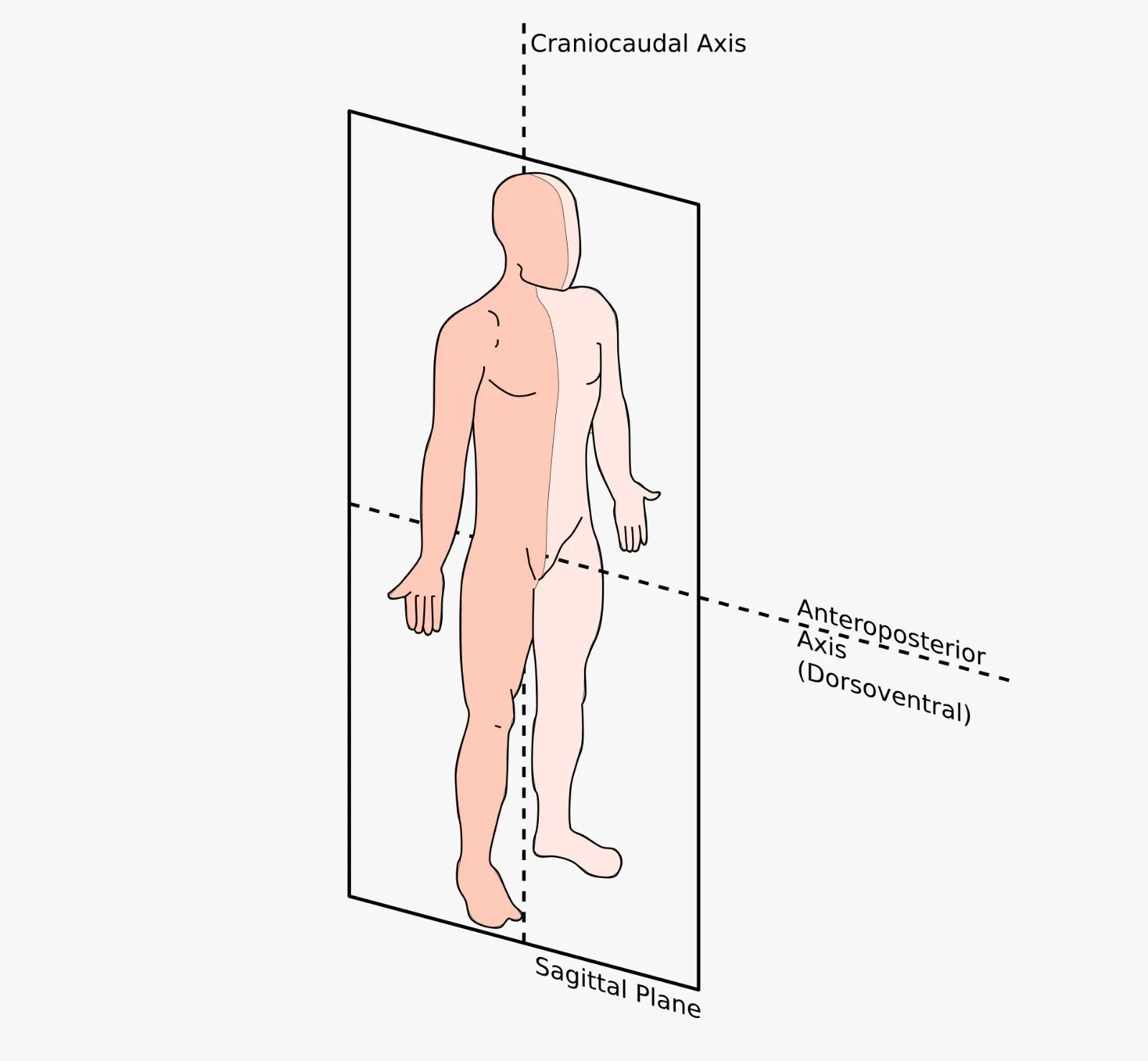
In anatomy, the sagittal plane is a vertical plane that runs from the anterior (front) to the posterior (back) of the body, dividing it into left and right portions. The term is derived from the Latin word sagitta, meaning "arrow," referring to the path an arrow would take when shot through the body from front to back. This plane is fundamental in describing movements and anatomical relationships.
Types of Sagittal Planes:
1. Midsagittal (Median) Plane
- Also called the median plane or midline
- Passes vertically through the center of the body
- Divides the body into equal left and right halves
- Passes through midline structures such as the nose, umbilicus, and spinal cord
- Example: A sagittal MRI of the brain showing both hemispheres separated by the corpus callosum
2. Parasagittal Plane
- Also called a sagittal section (non-midline)
- Passes vertically through the body but parallel to the midline
- Divides the body into unequal left and right portions
- Can be positioned anywhere to the left or right of the median plane
- Example: A cut through the right kidney, separating it from the left kidney but not dividing the body equally
Clinical Note: The sagittal plane is crucial for understanding flexion and extension movements (e.g., nodding the head, bending the knee). In radiology, midsagittal views are essential for assessing spinal alignment and brain structures, while parasagittal views help visualize organs like the lungs and kidneys individually. Surgical approaches to the spine often utilize a parasagittal incision to avoid damaging critical midline structures.
Coronal Plane
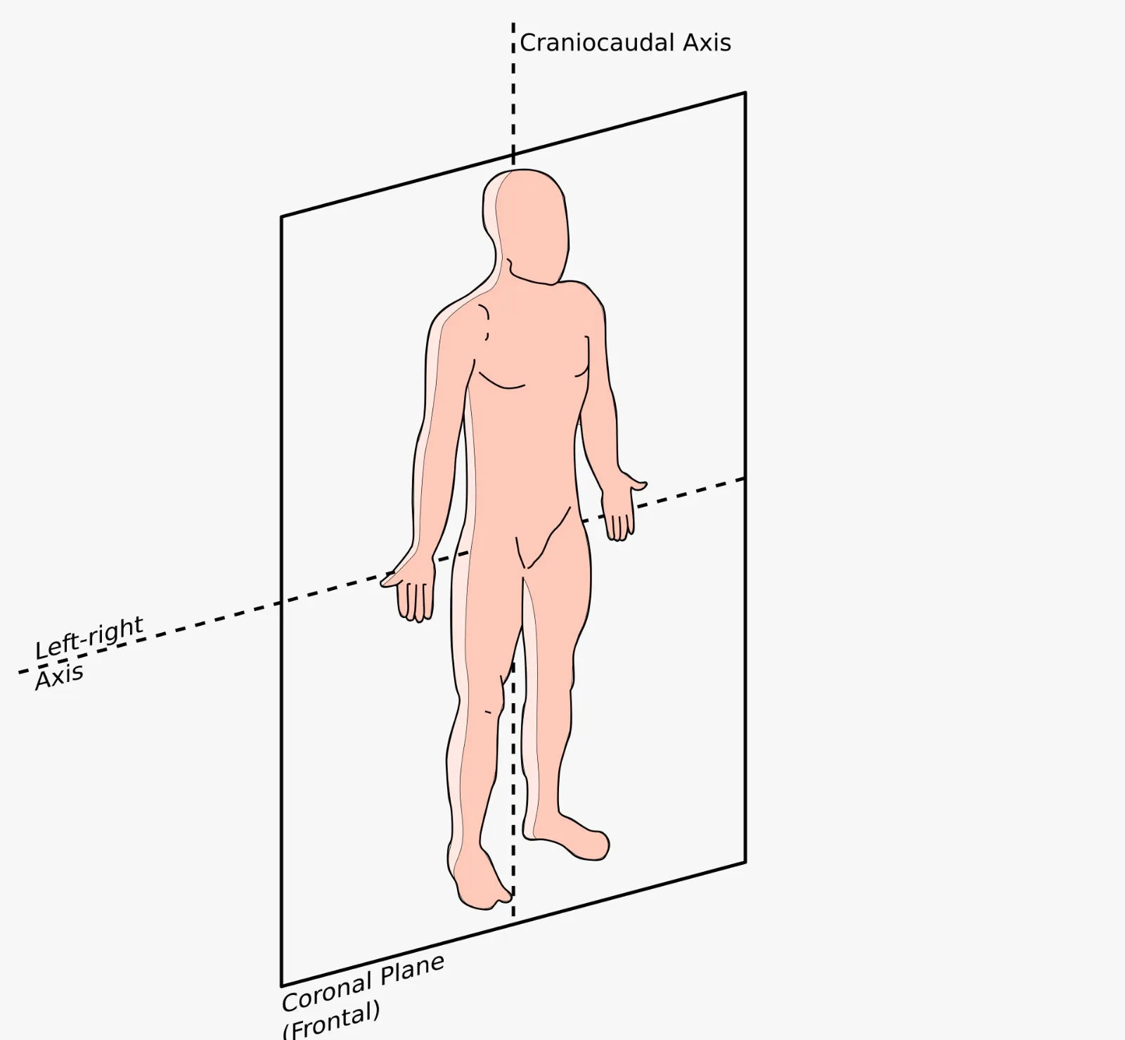
In anatomy, the coronal plane, also known as the frontal plane, is a vertical plane that runs from side to side (right to left), dividing the body into anterior (front) and posterior (back) sections. The term is derived from the Latin word corona, meaning "crown," as it passes through the coronal suture of the skull—the line where the frontal bone meets the parietal bones.
Key Features:
- Vertical orientation (perpendicular to the ground in anatomical position)
- Runs from side to side (lateral to lateral)
- Divides the body into anterior (front) and posterior (back) portions
- Any plane parallel to the coronal suture of the skull is considered a coronal plane
- Multiple coronal planes can exist at different positions along the longitudinal axis
Movements in the Coronal Plane:
- Abduction: Movement away from the midline (e.g., raising arm sideways)
- Adduction: Movement toward the midline (e.g., lowering arm to side)
- Lateral flexion: Bending the spine or neck to the side
- Eversion/Inversion: Turning the sole of the foot outward or inward
- Radial/Ulnar deviation: Side-to-side movement of the wrist
Clinical Note: The coronal plane is essential in medical imaging, particularly in coronal MRI and CT scans, which provide clear views of structures like the lungs, heart, and pelvic organs from front to back. In orthopedics, understanding this plane is crucial for assessing scoliosis (lateral curvature of the spine) and planning surgical corrections. The term "frontal plane" is often used interchangeably, especially in kinesiology and biomechanics.
Transverse Plane
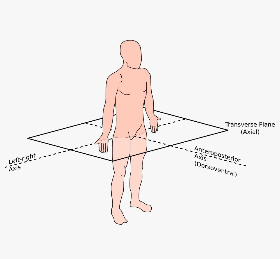
In anatomy, the transverse plane, also known as the horizontal plane or axial plane, is an imaginary plane that divides the body into superior (upper) and inferior (lower) portions. Unlike the sagittal and coronal planes which are vertical, the transverse plane is oriented horizontally, running parallel to the ground when the body is in the anatomical position. It is perpendicular to both the sagittal and coronal planes.
Key Features:
- Horizontal orientation (parallel to the ground)
- Divides the body into superior (cranial/upper) and inferior (caudal/lower) sections
- Perpendicular to both the sagittal and coronal planes
- Also called axial plane in radiology and medical imaging
- Multiple transverse planes can exist at different levels along the longitudinal axis (e.g., at T4 vertebra, L2 vertebra, mid-thigh)
Movements in the Transverse Plane:
- Rotation: Twisting of the head, spine, or limbs around their longitudinal axis
- Pronation/Supination: Rotation of the forearm (palm down/palm up)
- Medial (Internal) Rotation: Turning a limb toward the midline (e.g., rotating thigh inward)
- Lateral (External) Rotation: Turning a limb away from the midline (e.g., rotating thigh outward)
- Horizontal Abduction/Adduction: Moving the arm horizontally across the body (e.g., during a chest fly exercise)
Clinical Note: The transverse plane is the foundation of axial imaging—CT scans and MRI produce images in this plane, providing cross-sectional views of the body. This is invaluable for diagnosing pathologies in organs, blood vessels, and the spine. In surgery, understanding transverse plane anatomy is critical for procedures like spinal pedicle screw placement and joint arthroplasty. The term "axial" is preferred in radiology because images are taken along the axis of the body.
Please avoid hate, spam, or offensive content. Every comment is monitored, and we aim to keep this space respectful and safe for all users.