
Body Cavities
A body cavity is any fluid-filled space or compartment in the body that houses and protects internal organs (viscera). These cavities allow organs to move, expand, and function without distorting surrounding tissues. The body maintains its internal organization by means of membranes, sheaths, and other structures that separate these compartments. The two largest human body cavities are the dorsal (posterior) cavity and the ventral (anterior) cavity.
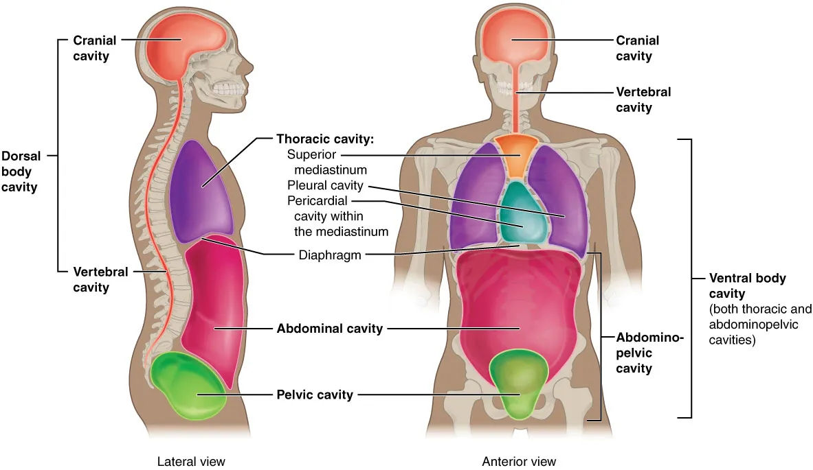
1. Dorsal Body Cavity (Posterior)
The dorsal cavity is located at the back of the body and protects the central nervous system. It is subdivided into two continuous cavities:
- Cranial cavity: A large, bean-shaped cavity formed by the skull that contains the brain. It is protected by the bones of the skull and cerebrospinal fluid.
- Spinal cavity (Vertebral canal): A long, narrow cavity within the vertebral column (spine) that contains the spinal cord. It is continuous with the cranial cavity.
Protective layers: The brain and spinal cord are enclosed by three membranes called the meninges (dura mater, arachnoid mater, and pia mater). Between these layers, cerebrospinal fluid circulates to cushion and protect neural structures.
Clinical note (Meningitis): Inflammation of the meninges, usually due to bacterial or viral infection, can lead to serious complications such as deafness, epilepsy, or cognitive deficits and is considered a medical emergency.
2. Ventral Body Cavity (Anterior)
The ventral cavity is located at the front of the trunk and contains the internal organs (viscera) of the respiratory, cardiovascular, digestive, urinary, and reproductive systems. It allows for significant changes in organ size and shape during functions such as breathing, digestion, and pregnancy. It is subdivided into the thoracic cavity and the abdominopelvic cavity, separated by the diaphragm.
2.1 Thoracic Cavity (Chest)
Enclosed by the rib cage and separated from the abdominopelvic cavity by the diaphragm. It contains:
- Pleural cavities (2): Each surrounds a lung and is lined by the pleura.
- Pericardial cavity: Surrounds the heart and is lined by the pericardium.
- Mediastinum: The central portion of the thoracic cavity between the lungs; contains the heart, esophagus, trachea, thymus, and major blood vessels.
2.2 Abdominopelvic Cavity
The largest cavity in the body, occupying the lower half of the trunk. Although no membrane physically divides it, it is subdivided for anatomical and clinical purposes:
Abdominal Cavity (Upper portion):
- Contains digestive organs: stomach, small and large intestines, liver, gallbladder, pancreas.
- Also contains spleen and kidneys (though kidneys are retroperitoneal, behind the peritoneum).
- Bounded superiorly by the diaphragm, anteriorly by abdominal muscles, posteriorly by lumbar vertebrae.
Pelvic Cavity (Lower portion):
- Funnel-shaped and continuous with the abdominal cavity.
- Contains: urinary bladder, reproductive organs (uterus, ovaries in females; prostate, seminal vesicles in males), rectum.
- Bounded by pubic bones, sacrum, coccyx, and pelvic floor muscles.
Abdominopelvic Regions and Quadrants:
To communicate precisely about locations (e.g., pain or masses), healthcare providers divide the abdominopelvic cavity into:
- Nine regions: Using two horizontal lines (subcostal and transtubercular) and two vertical lines (midclavicular lines).
- Four quadrants: Using one horizontal and one vertical line intersecting at the umbilicus (belly button).
Serous Membranes (Serosa)
Serous membranes are thin, double-layered membranes that line the ventral body cavities and cover the organs within them. They secrete a thin, lubricating serous fluid that reduces friction between organs and cavity walls during movement (e.g., heart beating, lungs inflating, intestines moving).
Two layers of serous membranes:
- Parietal layer: Lines the internal surface of the body wall.
- Visceral layer: Covers the external surface of the organs (viscera).
Between these layers is a potential space called the serous cavity, containing a thin film of serous fluid.
Three Major Serous Membranes:
- Pleura: Surrounds the lungs (pleural cavity).
- Pericardium: Surrounds the heart (pericardial cavity).
- Peritoneum: Surrounds abdominal and pelvic organs (peritoneal cavity).
Clinical note: Inflammation of these membranes (e.g., peritonitis, pleurisy, pericarditis) causes severe pain due to friction between the inflamed layers and can be life-threatening.
Summary Table: Body Cavities and Membranes
| Major Cavity | Subdivision | Principal Contents | Serous Membrane |
|---|---|---|---|
| Dorsal | Cranial cavity | Brain | Meninges |
| Dorsal | Spinal cavity | Spinal cord | Meninges |
| Ventral | Thoracic cavity (Pleural cavities) | Lungs | Pleura |
| Ventral | Thoracic cavity (Pericardial cavity) | Heart | Pericardium |
| Ventral | Thoracic cavity (Mediastinum) | Heart, esophagus, trachea, thymus, great vessels | — |
| Ventral | Abdominal cavity | Digestive organs, spleen, kidneys | Peritoneum |
| Ventral | Pelvic cavity | Bladder, reproductive organs, rectum | Peritoneum |
Clinical Note Summary: Body cavities are essential for organ protection and function. Meningitis (inflammation of meninges) affects the dorsal cavity. Pleurisy, pericarditis, and peritonitis are inflammatory conditions of the serous membranes lining the ventral cavity. The abdominopelvic cavity's division into quadrants/regions helps localize pathologies (e.g., appendicitis pain in right lower quadrant). During pregnancy, the ventral cavity accommodates fetal growth because organs can expand without disrupting nearby structures.
Please avoid hate, spam, or offensive content. Every comment is monitored, and we aim to keep this space respectful and safe for all users.