
Anatomical Position
When a person is standing straight with eyes looking forwards, both arms by the side of body, palms facing forwards, both feet together, the position is anatomical position.
Features
- Standing upright
- Head & eyes facing forward
- Arms by side
- Palms facing forward
- Feet flat and forward
All anatomical terms are described based on this position.
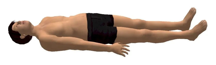
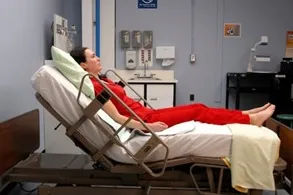
Supine Position
In anatomy, the supine position is a position of the body in which a person is lying horizontally on their back, facing upward. The face is directed vertically toward the ceiling, and the spine is aligned horizontally. This is considered the opposite of the prone position (lying face down).
Features
- Lying horizontally on the back
- Head and eyes facing upward (forward relative to horizon)
- Arms by the side of the body (palms may face up or toward body)
- Legs extended, feet typically pointing slightly outward
- Spine in a neutral, horizontal alignment
Clinical Note: While standard for surgery and exams, the supine position can cause airway obstruction by the tongue or, in late pregnancy, compress the inferior vena cava leading to hypotension. Prolonged immobilization in this position risks pressure sores on the sacrum and heels.
Prone Position
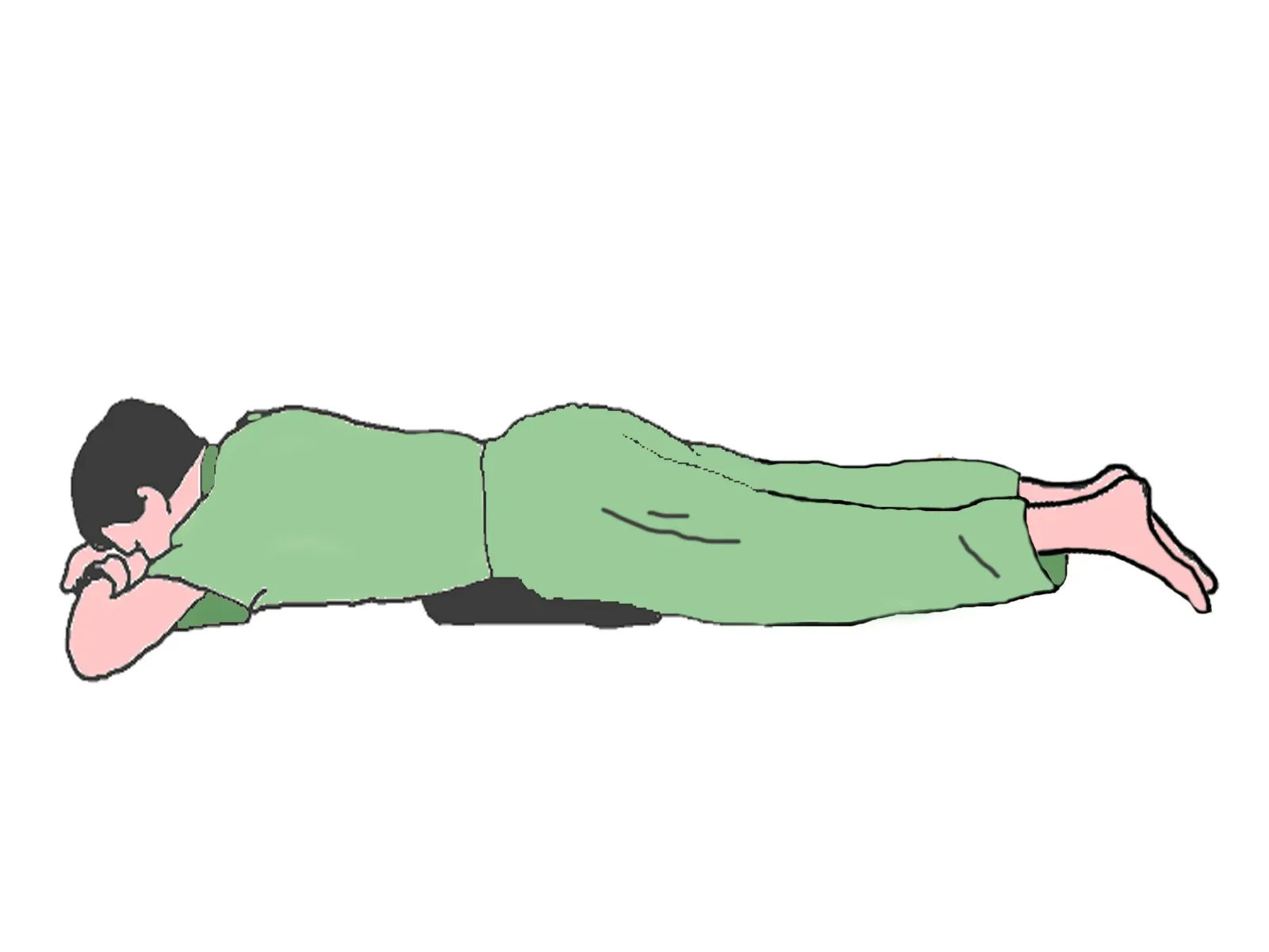
In anatomy, the prone position is a position of the body in which a person is lying horizontally on their stomach, facing downward. The face is typically turned to one side to allow for breathing, and the back is the most superficial aspect of the body. This is the direct opposite of the supine position (lying on the back).
Features
- Lying horizontally on the stomach (anterior surface)
- Head turned to one side (typically) to maintain airway
- Arms may be positioned beside the head or alongside the body
- Legs extended, feet in neutral position
- Spine maintains its natural curves under gravity
Clinical Note: The prone position is essential for surgeries involving the posterior body (spine, back). It is also used therapeutically in patients with Acute Respiratory Distress Syndrome (ARDS) to improve oxygenation (prone positioning). However, careful padding is required to avoid pressure on the eyes, breasts, and genitals, and to maintain neck alignment.
Lateral Position
In anatomy, the lateral position is a position of the body in which a person is lying on their side. It is also commonly referred to as the side-lying position. When specified as right lateral, the person lies on their right side; left lateral means lying on the left side. This position is frequently used in surgery, medical imaging, and as a recovery position.
Features
- Lying horizontally on the side (right or left)
- Head supported in a neutral position, aligned with the spine
- Inferior arm (down side) may be positioned forward or under the head for support
- Superior arm (up side) is typically positioned forward or on a pillow
- Legs may be staggered with the top leg flexed over a pillow to stabilize the pelvis
Clinical Note: The lateral position is the basis of the recovery position used for unconscious patients to maintain a clear airway and prevent aspiration. In surgery (e.g., hip or thoracic procedures), it provides access to lateral structures, but requires careful padding of pressure points like the ear, shoulder, and greater trochanter, and an axillary roll to protect the neurovascular bundle of the dependent arm.
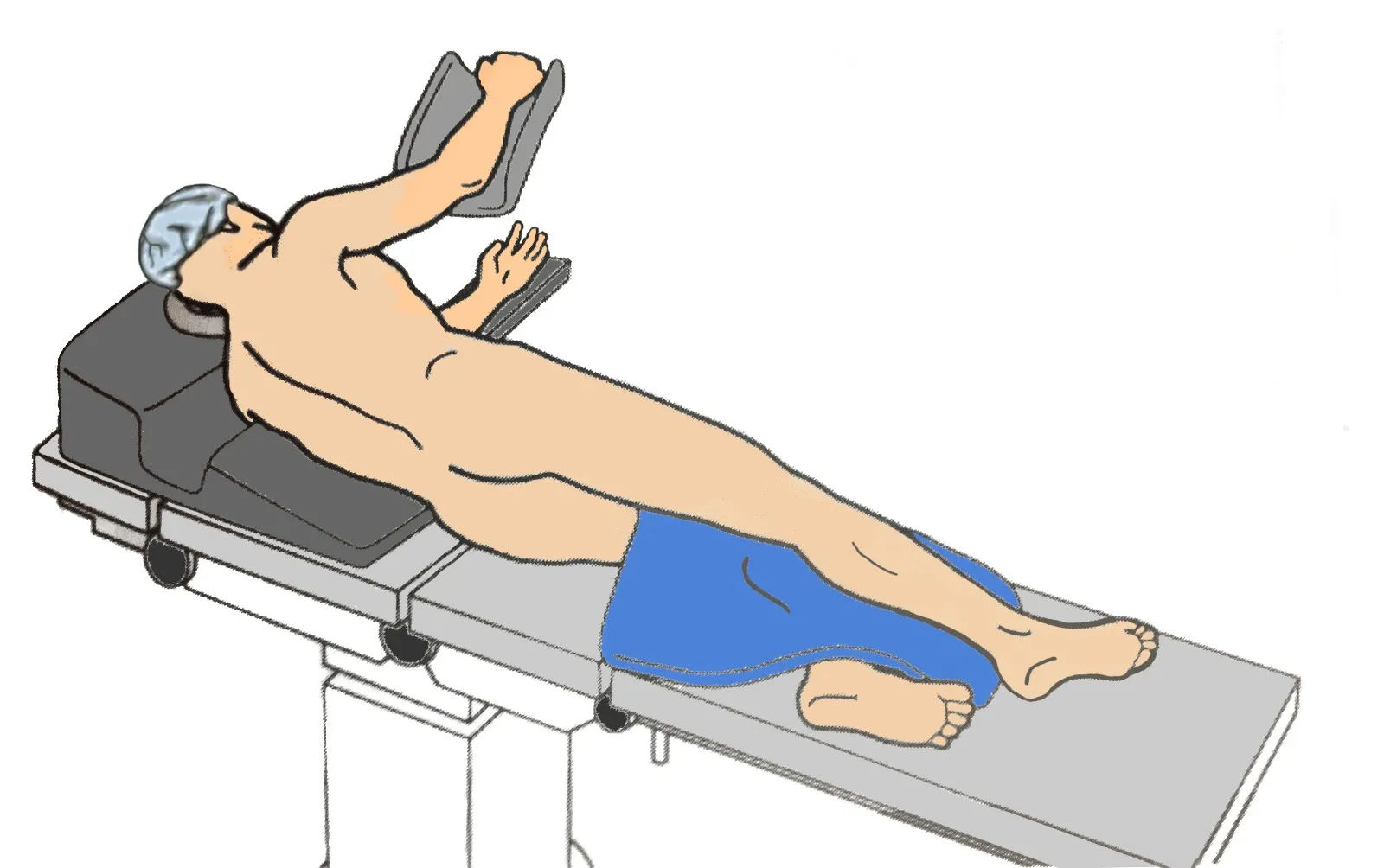
Lithotomy Position
In anatomy and surgery, the lithotomy position is a variation of the supine position in which the individual lies on their back with hips and knees fully flexed and legs elevated and supported in stirrups. The name is derived from the Greek words lithos (stone) and tomia (cut), as it was historically used for lithotomy—the surgical removal of bladder stones.
Features
- Lying on the back (supine) with buttocks positioned at the edge of the table
- Hips flexed between 80-100 degrees (or more, depending on variation)
- Knees flexed with lower legs supported by stirrups
- Thighs abducted (separated) to allow access to the perineum
- Arms may be tucked at sides or extended on arm boards
Clinical Note: The lithotomy position provides excellent surgical access for gynecological, urological, and colorectal procedures (e.g., childbirth, hysterectomy, prostate surgery). However, it carries significant risks: prolonged positioning can cause compartment syndrome of the lower legs, common peroneal nerve injury (foot drop), and positioning-related hip injuries. Legs must be lifted and lowered simultaneously to avoid torsion on the lumbar spine.
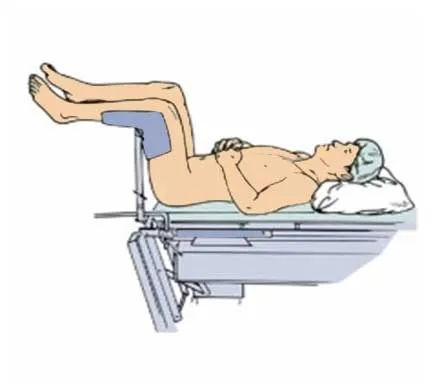
Fowler's Position
In anatomy and clinical medicine, Fowler's position is a standard patient position in which the individual is seated in a semi-upright posture at an angle between 45 and 60 degrees. Named after surgical pioneer George Ryerson Fowler, this position is achieved by elevating the head of the bed while optionally flexing the knees slightly to prevent the patient from sliding downward. Variations include Low Fowler's (15-30 degrees), Semi-Fowler's (30-45 degrees), and High Fowler's (60-90 degrees).
Features:
- Sitting upright with head elevated between 45-90 degrees (depending on variation)
- Knees slightly flexed (usually 15 degrees) to support the legs and prevent sliding
- Feet supported by a footboard to prevent foot drop and maintain position
- Arms may rest on pillows or bedside tables for support
- Head and neck maintained in neutral alignment with the spine
Clinical Note: Fowler's position is essential for patients with respiratory distress (e.g., COPD, pneumonia) as it allows for maximum chest expansion and diaphragmatic descent, improving oxygenation. It is also used for feeding, swallowing evaluations, and intracranial pressure management. However, prolonged positioning requires pressure relief on the sacrum and heels, and caution is needed in hypotensive patients as the upright posture can reduce cerebral perfusion.
Anatomical Planes
Sagittal Plane
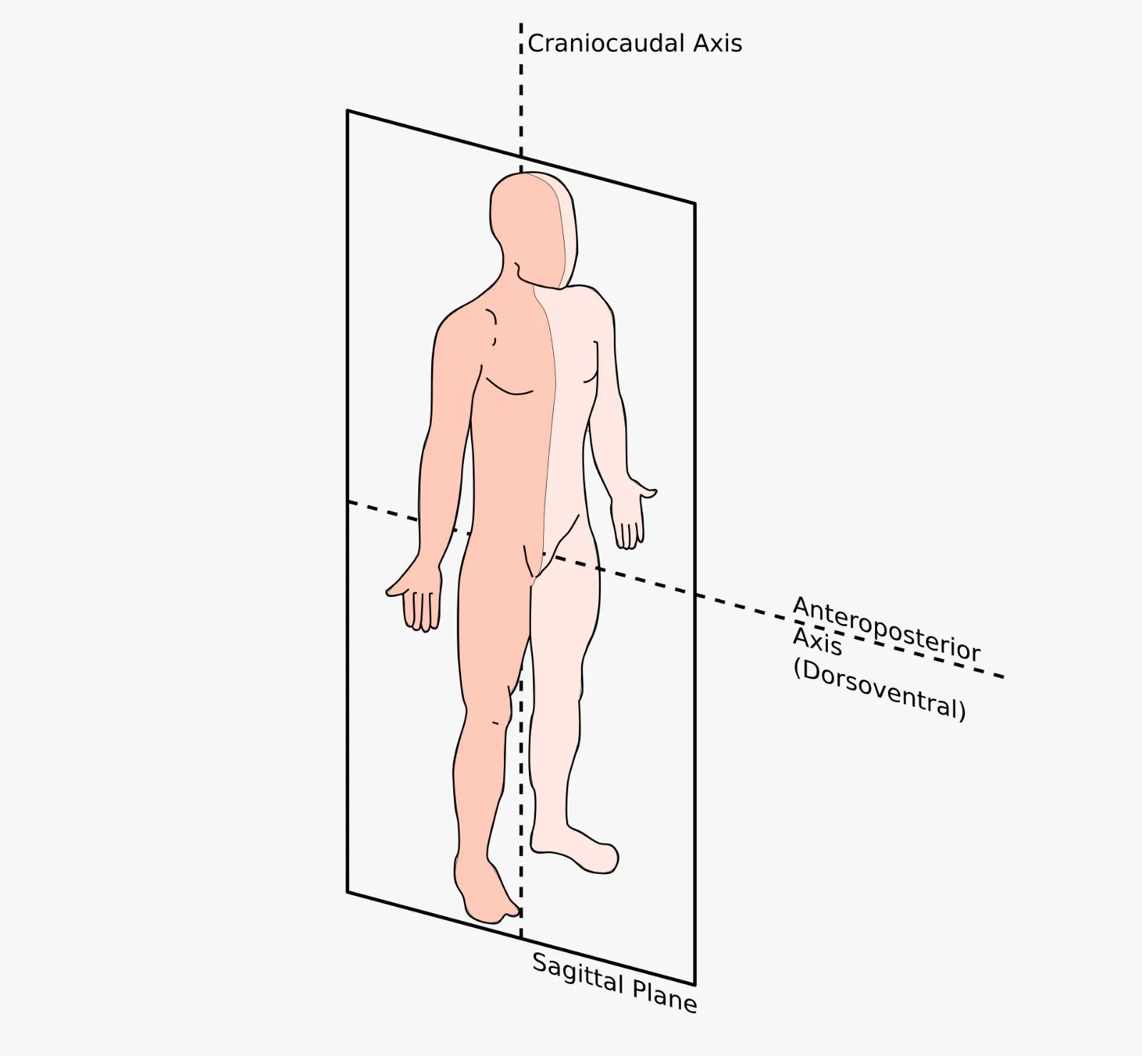
In anatomy, the sagittal plane is a vertical plane that runs from the anterior (front) to the posterior (back) of the body, dividing it into left and right portions. The term is derived from the Latin word sagitta, meaning "arrow," referring to the path an arrow would take when shot through the body from front to back. This plane is fundamental in describing movements and anatomical relationships.
Types of Sagittal Planes:
1. Midsagittal (Median) Plane
- Also called the median plane or midline
- Passes vertically through the center of the body
- Divides the body into equal left and right halves
- Passes through midline structures such as the nose, umbilicus, and spinal cord
- Example: A sagittal MRI of the brain showing both hemispheres separated by the corpus callosum
2. Parasagittal Plane
- Also called a sagittal section (non-midline)
- Passes vertically through the body but parallel to the midline
- Divides the body into unequal left and right portions
- Can be positioned anywhere to the left or right of the median plane
- Example: A cut through the right kidney, separating it from the left kidney but not dividing the body equally
Clinical Note: The sagittal plane is crucial for understanding flexion and extension movements (e.g., nodding the head, bending the knee). In radiology, midsagittal views are essential for assessing spinal alignment and brain structures, while parasagittal views help visualize organs like the lungs and kidneys individually. Surgical approaches to the spine often utilize a parasagittal incision to avoid damaging critical midline structures.
Coronal Plane
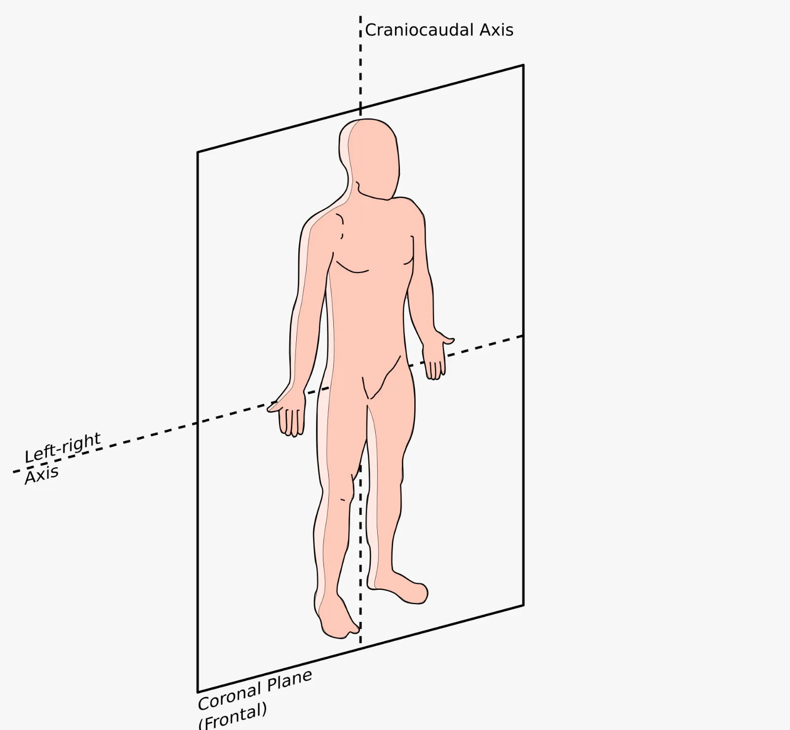
In anatomy, the coronal plane, also known as the frontal plane, is a vertical plane that runs from side to side (right to left), dividing the body into anterior (front) and posterior (back) sections. The term is derived from the Latin word corona, meaning "crown," as it passes through the coronal suture of the skull—the line where the frontal bone meets the parietal bones.
Key Features:
- Vertical orientation (perpendicular to the ground in anatomical position)
- Runs from side to side (lateral to lateral)
- Divides the body into anterior (front) and posterior (back) portions
- Any plane parallel to the coronal suture of the skull is considered a coronal plane
- Multiple coronal planes can exist at different positions along the longitudinal axis
Movements in the Coronal Plane:
- Abduction: Movement away from the midline (e.g., raising arm sideways)
- Adduction: Movement toward the midline (e.g., lowering arm to side)
- Lateral flexion: Bending the spine or neck to the side
- Eversion/Inversion: Turning the sole of the foot outward or inward
- Radial/Ulnar deviation: Side-to-side movement of the wrist
Clinical Note: The coronal plane is essential in medical imaging, particularly in coronal MRI and CT scans, which provide clear views of structures like the lungs, heart, and pelvic organs from front to back. In orthopedics, understanding this plane is crucial for assessing scoliosis (lateral curvature of the spine) and planning surgical corrections. The term "frontal plane" is often used interchangeably, especially in kinesiology and biomechanics.
Transverse Plane
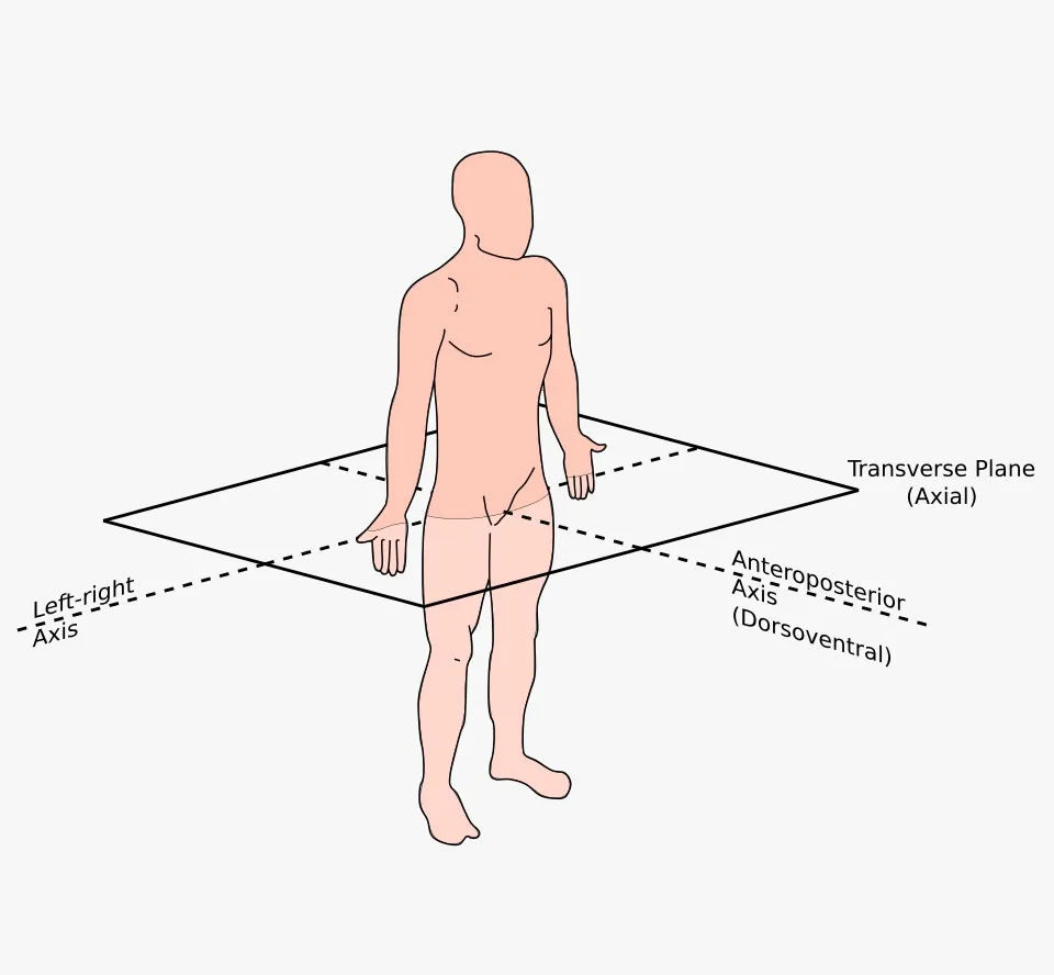
In anatomy, the transverse plane, also known as the horizontal plane or axial plane, is an imaginary plane that divides the body into superior (upper) and inferior (lower) portions. Unlike the sagittal and coronal planes which are vertical, the transverse plane is oriented horizontally, running parallel to the ground when the body is in the anatomical position. It is perpendicular to both the sagittal and coronal planes.
Key Features:
- Horizontal orientation (parallel to the ground)
- Divides the body into superior (cranial/upper) and inferior (caudal/lower) sections
- Perpendicular to both the sagittal and coronal planes
- Also called axial plane in radiology and medical imaging
- Multiple transverse planes can exist at different levels along the longitudinal axis (e.g., at T4 vertebra, L2 vertebra, mid-thigh)
Movements in the Transverse Plane:
- Rotation: Twisting of the head, spine, or limbs around their longitudinal axis
- Pronation/Supination: Rotation of the forearm (palm down/palm up)
- Medial (Internal) Rotation: Turning a limb toward the midline (e.g., rotating thigh inward)
- Lateral (External) Rotation: Turning a limb away from the midline (e.g., rotating thigh outward)
- Horizontal Abduction/Adduction: Moving the arm horizontally across the body (e.g., during a chest fly exercise)
Clinical Note: The transverse plane is the foundation of axial imaging—CT scans and MRI produce images in this plane, providing cross-sectional views of the body. This is invaluable for diagnosing pathologies in organs, blood vessels, and the spine. In surgery, understanding transverse plane anatomy is critical for procedures like spinal pedicle screw placement and joint arthroplasty. The term "axial" is preferred in radiology because images are taken along the axis of the body.
Levels of Organization
The human body is structured in a highly organized hierarchy, ranging from the simplest chemical building blocks to the complex, living organism. This concept, known as the levels of structural organization, demonstrates how smaller components assemble to form larger, more functional units. Understanding this hierarchy is fundamental to studying anatomy and physiology, as each level builds upon the previous one.
The Six Levels (Simplest to Most Complex):
1. Chemical Level (Biochemical Foundation of Life)
The chemical level is the smallest and most fundamental level of structural organization. It forms the foundation for all biological processes that sustain life. This level concerns the interaction of atoms and molecules, which ultimately determines the structure and function of cells, tissues, and organs.
It includes:
- Atoms: The basic units of matter
- Molecules: Combinations of atoms held by chemical bonds
- Macromolecules: Large, complex molecules essential for life
Atoms: The Building Blocks
Atoms are the smallest units of matter that retain the properties of an element and participate in chemical reactions. The human body is composed of approximately 24 essential elements, with four making up about 96% of body mass.
Major Elements in the Human Body (by percentage):
- Oxygen (O) - 65%: Essential for cellular respiration and water formation
- Carbon (C) - 18.5%: The backbone of organic molecules; forms four stable bonds
- Hydrogen (H) - 9.5%: Component of water and organic compounds; proton donor in reactions
- Nitrogen (N) - 3.2%: Fundamental component of proteins and nucleic acids (DNA/RNA)
Lesser but Essential Elements (Trace Elements):
- Calcium (Ca): Bone structure, muscle contraction, nerve signaling
- Phosphorus (P): ATP energy currency, nucleic acids, bone matrix
- Potassium (K): Membrane potential, nerve impulses
- Sulfur (S): Protein structure (disulfide bridges), enzyme function
- Sodium (Na): Fluid balance, nerve transmission
- Chlorine (Cl): Osmotic balance, stomach acid (HCl)
- Magnesium (Mg): Enzyme cofactor, ATP stabilization
- Iron (Fe): Hemoglobin oxygen transport, electron transport chain
- Iodine (I): Thyroid hormone synthesis
Molecules and Compounds
Atoms combine through chemical bonds (covalent, ionic, or hydrogen) to form molecules and compounds. These range from simple inorganic molecules to complex macromolecules.
Inorganic Molecules:
- Water (H₂O): The universal solvent; makes up 50-70% of body mass; crucial for temperature regulation, transport, and hydrolysis reactions
- Oxygen (O₂): Required for cellular respiration and ATP production
- Carbon dioxide (CO₂): Waste product of metabolism; regulates blood pH
- Electrolytes (Na⁺, K⁺, Ca²⁺, Cl⁻, HCO₃⁻): Ions essential for nerve conduction, muscle contraction, and fluid balance
Organic Molecules (Carbon-based):
Carbohydrates
- Monosaccharides: Glucose (C₆H₁₂O₆) - primary cellular fuel
- Disaccharides: Sucrose, lactose
- Polysaccharides: Glycogen (energy storage in liver/muscles)
Lipids (Hydrophobic molecules)
- Triglycerides (fats): Long-term energy storage, insulation
- Phospholipids: Form cell membranes (bilayer structure)
- Steroids: Cholesterol (membrane fluidity), hormones (estrogen, testosterone)
Proteins (Polypeptides)
- Composed of amino acid chains folded into 3D structures
- Functions: Enzymes (catalysis), structural (collagen), transport (hemoglobin), defense (antibodies), regulation (hormones)
- Example: Hemoglobin carries O₂ in red blood cells
Nucleic Acids
- DNA (Deoxyribonucleic acid): Double helix; stores genetic information; composed of nucleotides (A, T, G, C)
- RNA (Ribonucleic acid): Single-stranded; involved in protein synthesis (mRNA, tRNA, rRNA)
- ATP (Adenosine triphosphate): The energy currency of the cell; captures and transfers chemical energy
Biochemical Reactions and Processes
The chemical level is not static—it is a dynamic environment of continuous reactions:
- Metabolism: Sum of all chemical reactions (catabolism breaks down, anabolism builds up)
- Enzymatic reactions: Proteins catalyze specific reactions (e.g., digestive enzymes break down food)
- Oxidation-reduction (redox): Electron transfer reactions essential for energy production
- Acid-base balance: pH regulation through buffer systems (bicarbonate, phosphate, proteins)
Clinical Correlation: When the Chemical Level Fails
- Electrolyte imbalance: Abnormal Na⁺ or K⁺ levels cause cardiac arrhythmias, muscle weakness, or coma
- Hypoxia: Insufficient O₂ at the chemical level leads to cellular death (e.g., stroke, myocardial infarction)
- Genetic mutations: Altered DNA sequence (chemical change) results in defective proteins (e.g., sickle cell anemia, cystic fibrosis)
- Diabetes mellitus: Disruption of glucose metabolism at the molecular level causes systemic complications
- Heavy metal toxicity: Lead or mercury binding to enzymes disrupts their function
Importance: Why the Chemical Level Matters
These chemicals and their interactions perform vital roles that sustain life:
- Energy production: ATP synthesis through cellular respiration (glucose + O₂ → CO₂ + H₂O + ATP)
- Building body structures: Collagen provides tensile strength to skin, bone, and tendons; calcium phosphate mineralizes bone matrix
- Regulating body functions: Hormones (chemical messengers) coordinate growth, metabolism, and reproduction; enzymes control reaction rates
- Information storage: DNA encodes hereditary information
- Protection: Antibodies recognize and neutralize pathogens
The chemical level reminds us that life, in its essence, is a beautifully orchestrated series of chemical interactions—disrupt one, and the entire organism feels the consequence.
2. Cellular Level (The Fundamental Unit of Life)
The cellular level is formed when molecules and macromolecules organize into specialized structures called cells. A cell is the smallest structural and functional unit of life capable of carrying out all life processes independently. While molecules alone are not alive, their precise organization within a cell creates the emergent property of life.
The human body is a complex society of approximately 37 trillion cells, each performing specific roles while communicating and cooperating to maintain homeostasis. Cells vary dramatically in size, shape, and function based on their specialized roles.
What Defines a Cell?
All human cells share certain fundamental characteristics:
- Plasma membrane: A phospholipid bilayer that separates internal contents from the external environment and regulates transport
- Cytoplasm: A gel-like substance containing organelles and cytoskeletal elements
- Genetic material (DNA): Instructions for protein synthesis and cell function
- Ribosomes: Sites of protein synthesis
- Metabolic capability: Ability to perform chemical reactions for energy production and maintenance
Major Organelles and Their Functions
Eukaryotic human cells contain specialized membrane-bound structures called organelles:
- Nucleus: Contains DNA organized into chromosomes; control center of the cell; site of transcription
- Mitochondria: "Powerhouses" of the cell; produce ATP through cellular respiration; contain their own DNA
- Endoplasmic Reticulum (ER):
- Rough ER: Studded with ribosomes; synthesizes proteins for secretion or membrane insertion
- Smooth ER: Lipid synthesis, detoxification, calcium storage
- Golgi apparatus: Modifies, sorts, and packages proteins for transport
- Lysosomes: Digestive organelles containing hydrolytic enzymes; break down waste and pathogens
- Peroxisomes: Detoxify harmful substances (e.g., hydrogen peroxide) and metabolize fatty acids
- Cytoskeleton: Network of protein filaments (microtubules, microfilaments, intermediate filaments) providing structure, movement, and transport
- Centrioles: Organize microtubules during cell division
Major Human Cell Types and Their Specializations
The human body contains over 200 distinct cell types. Here are major categories with detailed examples:
Nerve Cells (Neurons)
- Structure: Cell body (soma), dendrites (receive signals), and axon (transmits signals)
- Function: Generate and conduct electrical impulses (action potentials) for communication
- Special features: Synaptic terminals release neurotransmitters; some axons are myelinated for rapid conduction
- Location: Brain, spinal cord, peripheral nerves
- Clinical note: Neurons have limited regenerative capacity; damage is often permanent (spinal cord injury, stroke)
Muscle Cells (Myocytes)
- Three types:
- Skeletal muscle: Long, multinucleated, striated; voluntary movement; attached to bones
- Cardiac muscle: Branched, uninucleated, striated; involuntary; intercalated discs for synchronized contraction
- Smooth muscle: Spindle-shaped, non-striated; involuntary; surrounds hollow organs (blood vessels, intestines, uterus)
- Function: Contraction generates force for movement, circulation, and peristalsis
- Special features: Rich in actin and myosin filaments; abundant mitochondria for energy
Blood Cells (Hematopoietic Lineage)
- Red blood cells (Erythrocytes):
- Biconcave discs without nucleus or organelles
- Contain hemoglobin for oxygen transport
- Approximately 25 trillion in human body (most abundant cell type)
- Lifespan ~120 days; produced in bone marrow
- White blood cells (Leukocytes):
- Neutrophils: Phagocytic; first responders to bacterial infection
- Lymphocytes: T cells (cellular immunity), B cells (antibody production)
- Monocytes: Become macrophages; phagocytose debris and pathogens
- Eosinophils: Combat parasites; involved in allergic responses
- Basophils: Release histamine in inflammation
- Platelets (Thrombocytes): Cell fragments essential for blood clotting
Epithelial Cells
- Structure: Closely packed cells with tight junctions; form barriers
- Functions: Protection, absorption, secretion, sensation
- Types by shape: Squamous (flat), cuboidal (cube-shaped), columnar (tall)
- Types by layers: Simple (single layer), stratified (multiple layers), pseudostratified (appears layered)
- Examples: Skin epidermis (stratified squamous); intestinal lining (simple columnar with microvilli); lung alveoli (simple squamous for gas exchange)
Connective Tissue Cells
- Fibroblasts: Secrete extracellular matrix (collagen, elastin)
- Adipocytes (fat cells): Store lipids for energy and insulation
- Osteocytes: Mature bone cells; maintain bone matrix
- Chondrocytes: Cartilage cells; maintain cartilage matrix
- Mast cells: Release histamine in inflammatory responses
Cellular Physiology: What Cells Do
- Metabolism: Cells carry out thousands of chemical reactions simultaneously—catabolism (breaking down molecules for energy) and anabolism (synthesizing molecules for growth and repair). Mitochondria produce ATP; enzymes catalyze specific reactions.
- Responsiveness (Irritability): Cells detect and respond to changes in their environment. Examples: Neurons respond to neurotransmitters; immune cells respond to cytokines; muscle cells respond to electrical stimulation.
- Growth and Reproduction: Cells increase in size and divide through the cell cycle (mitosis for somatic cells; meiosis for gametes). Cell division replaces damaged cells and enables growth. Some cells (neurons, skeletal muscle) lose the ability to divide after maturation.
- Communication: Cells communicate via chemical signals (hormones, neurotransmitters, paracrine signals) and direct contact (gap junctions, cell adhesion molecules).
- Movement: Some cells move (sperm cells, immune cells migrating to infection sites); others move substances (cilia in respiratory tract move mucus).
- Transport: Cells regulate what enters and exits through selective permeability—passive transport (diffusion, osmosis) and active transport (pumps, vesicles).
- Protein Synthesis: DNA transcribed to RNA; RNA translated to proteins on ribosomes; proteins determine cell structure and function.
The Numbers: Scale of the Cellular Level
- Total cells: Approximately 37 trillion (3.7 × 10¹³) in an average adult human
- Cell size range: Most human cells are 10-100 micrometers (µm) in diameter (red blood cells ~7 µm; neurons can have meter-long axons)
- Cell turnover: Approximately 330 billion cells replaced daily (about 1% of body weight)
- Microbiome: Bacterial cells in/on human body roughly equal human cells (~38 trillion)
- Red blood cells alone: ~25 trillion; replaced at 2-3 million per second
Clinical Correlations: When the Cellular Level Fails
- Cancer: Uncontrolled cell division due to genetic mutations; cells ignore growth signals and avoid apoptosis (programmed cell death)
- Anemia: Insufficient red blood cells or hemoglobin; reduced oxygen-carrying capacity
- Leukemia: Cancer of white blood cells; abnormal proliferation in bone marrow
- Muscular dystrophy: Genetic disorder causing progressive muscle cell degeneration and weakness
- Neurodegenerative diseases: Alzheimer's, Parkinson's, ALS—progressive loss of specific neuron populations
- Mitochondrial diseases: Defects in mitochondrial DNA affect ATP production; affect high-energy tissues (brain, muscle)
- Sickle cell disease: Genetic mutation alters hemoglobin shape; red blood cells become crescent-shaped, clog vessels, and rupture
- Infections: Viruses (HIV, influenza) hijack cellular machinery; bacteria (tuberculosis) survive inside macrophages
Stem Cells: The Cellular Foundation of Repair
- Definition: Undifferentiated cells capable of self-renewal and differentiation into specialized cell types
- Types:
- Embryonic stem cells: Pluripotent; can become any cell type
- Adult stem cells: Multipotent; limited to specific lineages (e.g., hematopoietic stem cells produce blood cells)
- Induced pluripotent stem cells (iPSCs): Adult cells reprogrammed to embryonic-like state
- Clinical applications: Bone marrow transplants (hematopoietic stem cells); research into regenerative medicine for spinal cord injury, Parkinson's, diabetes
Key Points Summary
- Cells carry out metabolism: All chemical reactions sustaining life occur within cells—energy production, synthesis, waste processing
- Cells respond to stimuli: Cells detect and react to internal and external signals (hormones, pH changes, pathogens)
- Cells reproduce and maintain body functions: Cell division replaces damaged/dead cells; specialized cells perform specific tasks (contraction, secretion, conduction, defense)
- The human body contains about 37 trillion cells: A vast, coordinated society of specialized units working together
The cellular level bridges chemistry and physiology. Understanding cells means understanding how molecules organize into living systems—and how disruptions at this level manifest as disease.
3. Tissue Level
- Tissues are groups of similar cells that work together to perform a common function
- The four primary tissue types in the human body:
- Epithelial tissue: Covers surfaces, lines cavities, forms glands
- Connective tissue: Supports, protects, binds structures (e.g., bone, blood, fat)
- Muscle tissue: Generates force for movement
- Nervous tissue: Conducts electrical signals for communication
- Example: Simple squamous epithelium forms the lining of blood vessels
Read Tissues in detail later in this chapter.
4. Organ Level
- An organ is a structure composed of two or more tissue types working together to perform specific, complex functions
- Organs have recognizable shapes and specific locations in the body
- Most organs contain all four tissue types in varying proportions
- Example: The stomach contains epithelial tissue (lining), connective tissue (support), muscle tissue (churning), and nervous tissue (control)
Organ System Level
An organ system consists of multiple organs working together to perform major body functions.
Major organ systems include:
- Skeletal system
- Muscular system
- Nervous system
- Digestive system
- Respiratory system
- Circulatory system
- Urinary system
- Endocrine system
- Lymphatic system
- Reproductive system
- Integumentary system
Example:
Digestive system includes stomach, intestines, liver, pancreas.
6. Organism Level
- The highest level of organization
- Represents the total living individual
- All organ systems function together to maintain life, health, and homeostasis
- The organism level represents the sum total of all lower levels working in harmony
- Example: A living human being, where all 11 organ systems operate simultaneously and interdependently
Clinical Note: Disease processes can begin at any level and spread to others. For example, a genetic mutation (chemical level) may cause abnormal cell function (cellular level), leading to tissue damage (tissue level), organ failure (organ level), and systemic illness (organism level). This hierarchical understanding guides diagnosis—blood tests detect chemical/cellular changes, biopsies examine tissues, and imaging evaluates organs and systems.
Please see the flow chart of levels of organization to understand how the human body is structured from the chemical level to the organism level.
Please avoid hate, spam, or offensive content. Every comment is monitored, and we aim to keep this space respectful and safe for all users.