
Abdominopelvic Regions
The abdominopelvic cavity is the largest cavity in the body, containing organs of digestion, excretion, and reproduction. To precisely locate pain, tenderness, masses, or surgical landmarks, clinicians divide this area into 9 regions (clinical standard) or 4 quadrants (emergency/quick assessment). These divisions are based on imaginary planes passing through key bony landmarks.
Why Divide the Abdominopelvic Cavity?
- Pinpoint organ location: helps identify which organ might be involved (e.g., appendix in right iliac region).
- Describe pain accurately: “right hypochondriac pain” suggests gallbladder, while “epigastric burning” suggests stomach/ulcer.
- Guide physical examination: tenderness in a specific region directs differential diagnosis.
- Surgical planning: incisions and laparoscopic port placement respect region anatomy.
Clinical Relevance: Referred pain often crosses regions. For example, diaphragmatic irritation (spleen or liver) can refer to the shoulder (C3–C5 dermatomes). Always combine region assessment with deeper palpation and imaging.
The 9 Abdominopelvic Regions (Anatomical Grid)
The nine-region scheme is created by two horizontal planes and two vertical (midclavicular) planes. This grid transforms the abdomen into a precise topographic map used in anatomy education, radiology reports, and surgical notes.
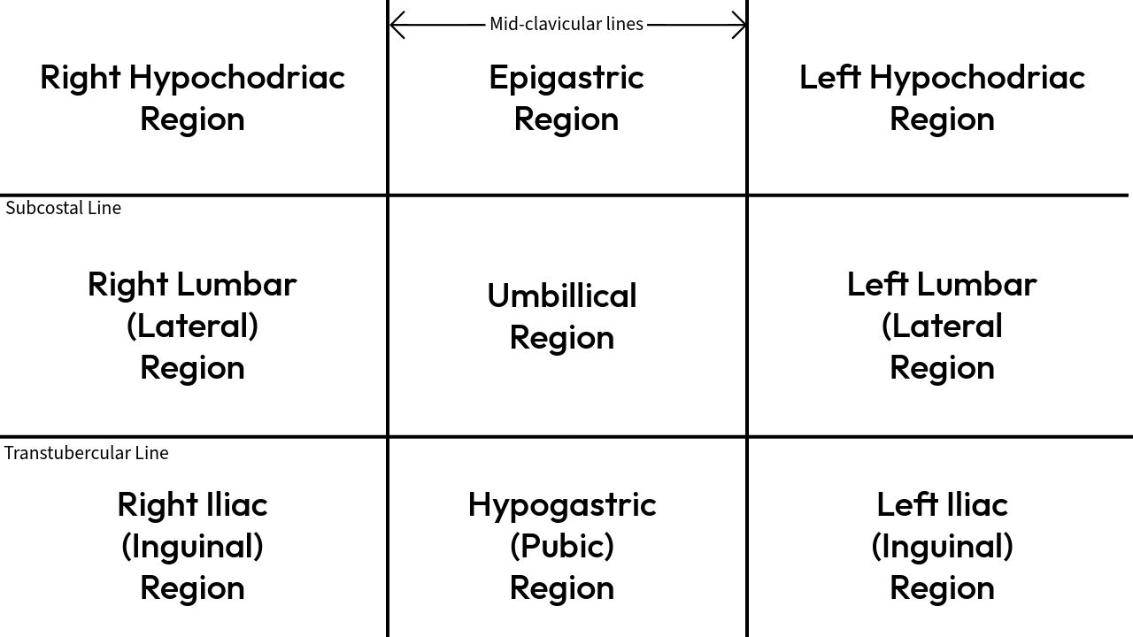
Landmarks Used to Create the 9 Regions
- Subcostal plane (superior horizontal): passes through the lowest point of the costal margin (10th costal cartilage) – approximates the level of L3 vertebra.
- Intertubercular plane (inferior horizontal): connects the two iliac tubercles (bony prominences of the anterior iliac crests) – roughly at L5 level.
- Two midclavicular lines (vertical): drawn vertically from the midpoint of each clavicle; they pass through the nipples in many individuals and traverse the inguinal ligament.
Top Row (Upper Abdomen)
- Right Hypochondriac Region: Right upper area, below the ribs. Contains right lobe of liver, gallbladder, right kidney (upper pole), hepatic flexure of colon.
- Epigastric Region: Central upper region, overlying the stomach. Houses stomach (body/pylorus), pancreas (body), left lobe of liver, duodenum, aorta, celiac trunk.
- Left Hypochondriac Region: Left upper area, below ribs. Contains spleen, stomach (fundus), left kidney (upper pole), splenic flexure of colon, tail of pancreas.
Tip: “Hypochondriac” = “below the cartilage” (rib cartilage). Epigastric = “above the stomach”.
Middle Row (Mid Abdomen – flank regions)
- Right Lumbar (Lateral) Region: Right side, lateral to umbilical region. Contains ascending colon, right kidney (lower pole), loops of small intestine.
- Umbilical Region: Central area around the umbilicus (navel). Contains transverse colon, greater omentum, small intestine (jejunum & ileum), abdominal aorta, inferior vena cava (IVC).
- Left Lumbar (Lateral) Region: Left side, lateral to umbilical region. Contains descending colon, left kidney (lower pole), jejunal loops.
Bottom Row (Lower Abdomen / Pelvic Regions)
- Right Iliac (Inguinal) Region: Lower right near the hip bone. Contains cecum, appendix, right ovary (female), right spermatic cord (male), right ureter. The appendix is most commonly located here – McBurney’s point.
- Hypogastric (Pubic) Region: Lower middle, above the pubic symphysis. Contains urinary bladder, uterus (female), rectum (lower part), sigmoid colon, prostate (male).
- Left Iliac (Inguinal) Region: Lower left near the hip bone. Contains sigmoid colon, left ovary (female), left spermatic cord (male), left ureter.
Memory aid (9 regions top to bottom, L to R): “Hippos Eat Lots, Right? Let’s Just Use Left Hypogastric.” But the systematic order: Right Hypochondriac, Epigastric, Left Hypochondriac, Right Lumbar, Umbilical, Left Lumbar, Right Iliac, Hypogastric, Left Iliac.
The 4 Quadrants (Rapid Clinical Assessment)
In emergency departments and bedside examinations, physicians often use the four-quadrant system. A single vertical midline (through the umbilicus) and a horizontal transumbilical line divide the abdomen into right upper (RUQ), left upper (LUQ), right lower (RLQ), and left lower (LLQ) quadrants. This method is fast but less precise than 9 regions.
Quadrants & Major Organ Relations
| Quadrant | Major Organs | Common Clinical Correlate |
|---|---|---|
| Right Upper Quadrant (RUQ) | Liver, Gallbladder, Duodenum, Head of pancreas, Right kidney | Cholecystitis, hepatitis, choledocholithiasis |
| Left Upper Quadrant (LUQ) | Spleen, Stomach, Left kidney, Tail of pancreas | Splenic rupture, gastritis, pancreatic tail tumor |
| Right Lower Quadrant (RLQ) | Appendix, Cecum, Right ovary/tube, Right ureter | Appendicitis, ovarian cyst, ectopic pregnancy, ureteric stone |
| Left Lower Quadrant (LLQ) | Sigmoid colon, Left ovary/tube, Left ureter | Diverticulitis, ovarian torsion, sigmoid volvulus |
Classic association: RLQ pain → think appendicitis; RUQ tenderness after fatty meal → cholecystitis; LLQ pain + fever → diverticulitis.
Comparison: 9 Regions vs. 4 Quadrants
- Precision: 9 regions offer detailed localization (e.g., epigastric vs hypogastric) – ideal for anatomy teaching and surgical reports. 4 quadrants trade detail for speed – ideal for trauma surveys.
- Usage: Quadrants dominate emergency medicine, nursing assessments, and general physical exams. Nine regions are preferred in radiology, gastroenterology, and medical school examinations.
- Overlap: Both systems complement each other. A clinician may first identify “RLQ pain” then refine to “right iliac region tenderness” for appendix localization.
Clinical pearl: Pain from the appendix may start in the umbilical region (referred visceral pain) and later localize to the right iliac region (parietal inflammation – McBurney’s sign). Similarly, pancreatitis often presents as epigastric pain radiating to the back, involving multiple regions. Understanding abdominopelvic regions improves diagnostic accuracy and communication between healthcare providers.
Region-by-Region Organ Summary Table
The table below condenses the nine regions with their primary organs. Use it as a rapid reference for exam or clinical reasoning.
| Region | Location / Boundaries | Principal Organs | Clinical Notes |
|---|---|---|---|
| Right Hypochondriac | Upper right, below ribs | Liver (right lobe), Gallbladder, Right kidney (upper pole), Hepatic flexure | Pain here suggests gallbladder issues (cholecystitis) or hepatitis |
| Epigastric | Upper middle, over stomach | Stomach, Pancreas (body), Duodenum, Left lobe of liver, Aorta | Burning pain → gastritis/ulcer; radiation to back → pancreatitis |
| Left Hypochondriac | Upper left, below ribs | Spleen, Stomach (fundus), Left kidney (upper pole), Tail of pancreas, Splenic flexure | Trauma + pain here → splenic rupture; referred shoulder pain (Kehr's sign) |
| Right Lumbar | Right side (lateral) | Ascending colon, Right kidney (lower pole), Small intestine loops | Flank pain + hematuria → renal colic (ureteric stone) |
| Umbilical | Central area around navel | Transverse colon, Small intestine (jejunum/ileum), Greater omentum, Aorta, IVC | Early appendicitis often starts as vague periumbilical pain |
| Left Lumbar | Left side (lateral) | Descending colon, Left kidney (lower pole), Jejunal loops | Diverticulitis is more common on left lower side, but left lumbar pain may occur with constipation |
| Right Iliac (Inguinal) | Lower right, near hip bone | Cecum, Appendix, Right ovary (F), Right spermatic cord (M), Right ureter | McBurney's point tenderness → appendicitis (most common surgical emergency) |
| Hypogastric (Pubic) | Lower middle, above pubis | Urinary bladder, Uterus (F), Sigmoid colon, Rectum, Prostate (M) | Suprapubic pain + frequency → cystitis; uterine tenderness → pelvic inflammatory disease |
| Left Iliac (Inguinal) | Lower left, near hip bone | Sigmoid colon, Left ovary (F), Left spermatic cord (M), Left ureter | LLQ pain + fever + change in bowel habits → diverticulitis (common in elderly) |
Note: "F" = female anatomy; "M" = male anatomy. Both sexes share most digestive and urinary organs.
Surface Anatomy & How to Find Each Region on a Patient
Palpating bony landmarks makes region identification consistent:
- Subcostal plane: Run your fingers along the lower edge of the rib cage (10th costal cartilage). This marks the upper border of the 9 regions (divides epigastric from umbilical and hypochondriac from lumbar).
- Intertubercular plane: Feel the iliac crests anteriorly; the most prominent bony bumps are the iliac tubercles (about 5 cm behind the ASIS). A horizontal line through these tubercles separates the lumbar regions from the iliac/hypogastric regions.
- Midclavicular lines: Located halfway between the sternal notch and the acromioclavicular joint. Drop vertically – they delineate the lateral borders of the epigastric/umbilical/hypogastric zones from the hypochondriac/lumbar/iliac zones.
Clinical note: The appendix lies at McBurney’s point – one-third of the distance from the right anterior superior iliac spine (ASIS) to the umbilicus; this corresponds to the center of the right iliac region.
Exam Tip: Many exam questions describe a patient with “right lower quadrant pain” and ask for the most likely diagnosis. However, if they ask for region name, the correct answer is “right iliac (inguinal) region.” Distinguish quadrant vs region terminology — the nine-region name should be used in anatomical contexts.
Clinical Syndromes & Common Region Associations
- Cholecystitis: Right hypochondriac pain, often radiating to right shoulder or scapula (referred via phrenic nerve). Murphy’s sign elicited under right costal margin.
- Appendicitis: Begins as vague periumbilical pain (referred visceral), then migrates to right iliac region (somatic pain). Tenderness at McBurney’s point.
- Diverticulitis: Typically left iliac region pain, fever, change in bowel habits. Most common in sigmoid colon.
- Peptic ulcer disease: Epigastric pain, often burning, relieved by food or antacids. Posterior penetrating ulcer may cause back pain.
- Renal colic: Severe flank (lumbar region) pain radiating to groin. Right or left lumbar region depending on stone side.
- Ectopic pregnancy: Lower abdominal pain (hypogastric or iliac region) with vaginal bleeding; life-threatening if ruptured.
- Pancreatitis: Epigastric pain radiating to back, often associated with nausea and elevated amylase/lipase. Epigastric tenderness may be severe.
Quick Mnemonic for the 9 Regions (Superior to Inferior, Left to Right)
Use the phrase: “Right Hypochondriac, Epigastric, Left Hypochondriac, Right Lumbar, Umbilical, Left Lumbar, Right Iliac, Hypogastric, Left Iliac.”
Another memory trick: “Harry’s Eggs Look Runny, Under Low Roofs, Henry’s Lunch.” But best: draw a 3x3 grid and label from patient’s right (your left) to left (your right). Practice on a partner using anatomical position.
Summary takeaway: The abdominopelvic nine-region system is the gold standard for detailed clinical anatomy. It converts vague abdominal complaints into topographical diagnoses. For time-critical scenarios like trauma or acute abdomen, the four-quadrant approach is fast and efficient. Mastering both ensures precise communication with surgeons, radiologists, and emergency colleagues.
Please avoid hate, spam, or offensive content. Every comment is monitored, and we aim to keep this space respectful and safe for all users.